Home › Unlabelled › Rib Cage Anatomy Muscles / Rib Cage Muscles And Tendons / Axial Muscles Of The ...
Rib Cage Anatomy Muscles / Rib Cage Muscles And Tendons / Axial Muscles Of The ...
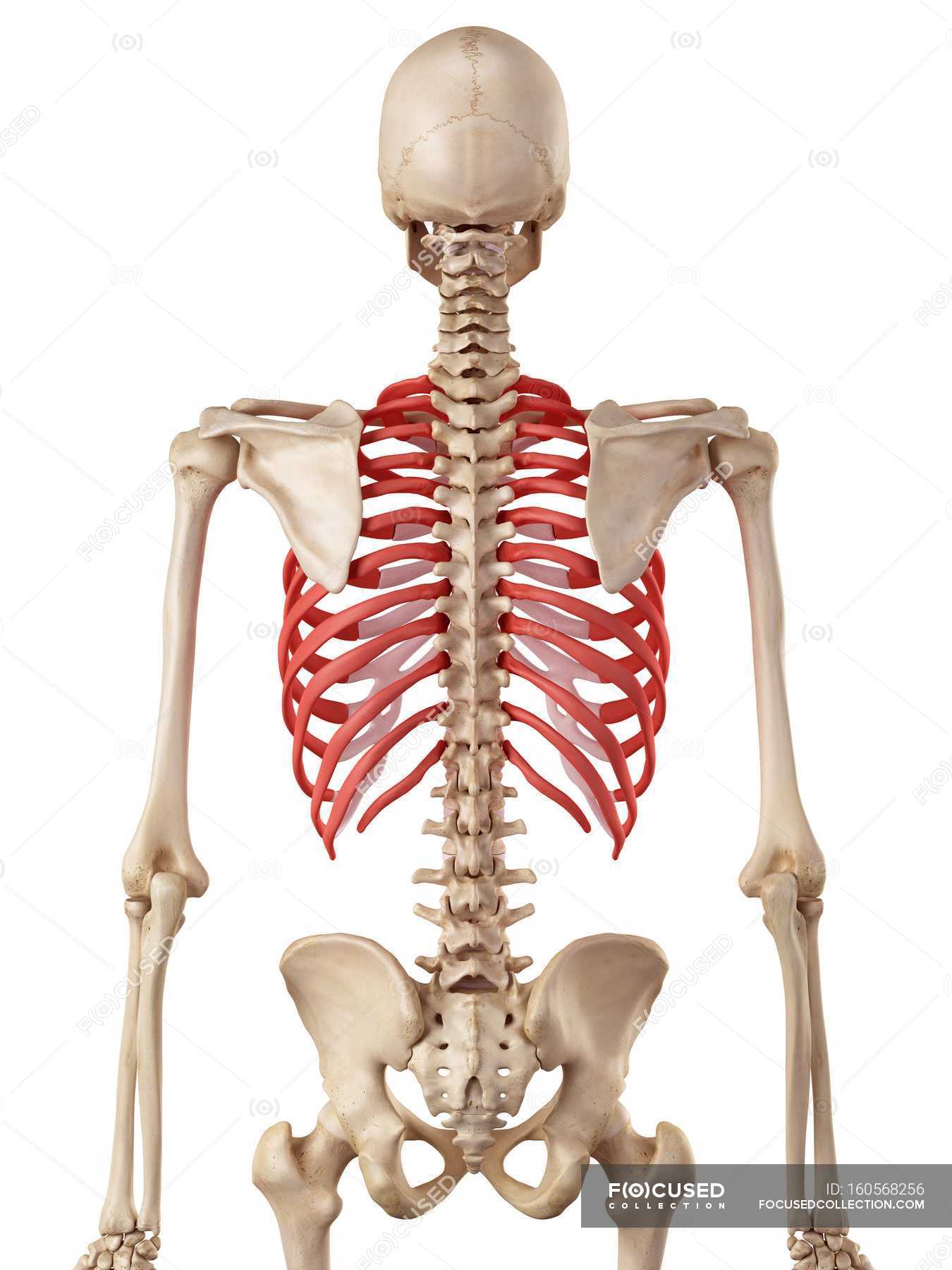
Rib Cage Anatomy Muscles / Rib Cage Muscles And Tendons / Axial Muscles Of The .... I also discussed the anatomy of false ribs, true ribs and floating ribs and the way they articulate with thoracic vertebrae and how they create the thoracic wall. Muscular system anatomy:muscles of the thoracic cage torso model description. • raise rib cage for inhaling & depresses rib cage for exhaling. The ribs are a set of twelve paired bones which form the protective 'cage' of the thorax. Volume rendering of a contrast enhanced thoracoabdominal ct scan.
The thoracic cage (rib cage) forms the thorax (chest) portion of the body. Muscle spasms located in the rib cage are often observed in people who strain or overwork their upper body muscles. Переглядів 46 тис.9 років тому. Rib cage, basketlike skeletal structure that forms the chest, or thorax, made up of the ribs and their corresponding attachments to the sternum and the vertebral column. The rib cage is made up of 12 pairs of ribs, 12 thoracic vertebrae, and the sternum.
Rib Cage Anatomy - Anatomy Color Coded Lungs Inside Rib ... from st.focusedcollection.com So what parts of the rib cage show up on the surface? The ribs are a set of twelve paired bones which form the protective 'cage' of the thorax. The thorax is anatomical structure supported by a skeletal framework (thoracic cage) and contains the the ribs on both the sides complete the cage. Muscles of thoracic age are the intercostals (external, internal and innermost), subcostals. Shaped somewhat like a cone, it is created by the individual ribs connecting to the spine above and to the sternum below. Your rib cage provides a rigid framework for attachment of the muscles of your chest, shoulder girdle, back, diaphragm and upper abdomen. This is a stereogram, to be viewed in crossview technique. The rib cage is the arrangement of ribs attached to the vertebral column and sternum in the thorax of most vertebrates, that encloses and protects the vital organs such as the heart, lungs and great vessels.
Your rib cage plays an important role in respiration, expanding and contracting as your respiratory muscles, including your diaphragm, work to help you breathe.
Muscles of the spine and rib cage | musculoskeletal key. Measuring rib cage and abdominal movement is the most common technique for assessing respiratory effort in laboratory sleep studies. Muscles that move the rib cage attach to the rib cage. So what parts of the rib cage show up on the surface? Muscles of the lower limb | anatomy model. Everyone has nice muscles in ct scanning! Structure of a typical rib: The rib cage is the arrangement of ribs attached to the vertebral column and sternum in the thorax of most vertebrates, that encloses and protects the vital organs such as the heart, lungs and great vessels. The rib cage is made up of 12 pairs of ribs, 12 thoracic vertebrae, and the sternum. These muscles may be located anteriorly, posteriorly, and/or laterally. The back end is wide and open. Human rib cage anatomy model. This is a stereogram, to be viewed in crossview technique.
Hd00:28skeletal system with muscles transparent animation. For a gesture drawing, that's good enough. When the upper arm is lifted away from the in poses such as this, you can see how the muscles change shape depending on the action of the arms and the placement of the rib cage and pelvis. • raise rib cage for inhaling & depresses rib cage for exhaling. The other attachment of these muscles is usually considered to be either superior or inferior to the rib attachment.
Learn Muscle Anatomy: Serratus Posterior Superior and ... from s-media-cache-ak0.pinimg.com The rib cage, shaped in a mild cone shape and more flexible than most bone sets, is made up of varying elements such as the thoracic vertebra, 12 equally paired ribs, costal cartilage, and held together anteriorly by the sternum. All muscles that are attached to the human rib cage have the inherent potential to cause a breathing action. 836 x 1024 jpeg 157kb. Muscles intercostal intercostals rib respiration muscle external internal anatomy ribcage ribs diagram pain costochondritis function physiology between thoracic breathing respiratory. Skeletal muscles attached to the rib cage: It consists of the 12 pairs of ribs with their costal cartilages and the sternum (figure in the anatomical position, the angles align with the medial border of the scapula. Volume rendering of a contrast enhanced thoracoabdominal ct scan. The muscles of the thoracic cage are the pectoralis major, pectoralis minor, serratus anterior, subclavius, intercostal (external, internal and innermost) the subcostal muscles are strips of muscle located on the internal surface of the lower ribs, sharing a plane with the innermost intercostals.
Various skeletal muscles are attached to the rib cage.
The rib cage is often simplified as an oval shape. The back end is wide and open. Muscles of the spine and rib cage | musculoskeletal key. Volume rendering of a contrast enhanced thoracoabdominal ct scan. Odd illustrations ribs bone thorax thorax bones floating ribs cartilaginous joint thoracic cage human chest anatomy spine draw vintage engraving anatomy. Muscles intercostal intercostals rib respiration muscle external internal anatomy ribcage ribs diagram pain costochondritis function physiology between thoracic breathing respiratory. So what parts of the rib cage show up on the surface? Each rib articulates posteriorly with the vertebral column. Muscles that helpful in expanding the thoracic cavity are called the inspiratory muscles because they help in inhalation, while those that compress the thoracic cavity are called expiratory. Hd00:28skeletal system with muscles transparent animation. 486 x 850 jpeg 55kb. Structure of a typical rib: 836 x 1024 jpeg 157kb.
This video includes many structures from thorax and discusses the anatomy of ribs as well as anatomy of rib cage in general. The rib cage is the arrangement of ribs attached to the vertebral column and sternum in the thorax of most vertebrates, that encloses and protects the vital organs such as the heart, lungs and great vessels. Skeletal muscles attached to the rib cage: Muscles of thoracic age are the intercostals (external, internal and innermost), subcostals. Structure of a typical rib:
MUSCULAR SYSTEM ANATOMY:Muscles of the Thoracic cage torso ... from i.ytimg.com All muscles that are attached to the human rib cage have the inherent potential to cause a breathing action. Some extend from above and draw the. The rib cage, shaped in a mild cone shape and more flexible than most bone sets, is made up of varying elements such as the thoracic vertebra, 12 equally paired ribs, costal cartilage, and held together anteriorly by the sternum. Everyone has nice muscles in ct scanning! • raise rib cage for inhaling & depresses rib cage for exhaling. It consists of the 12 pairs of ribs with their costal cartilages and the sternum (figure in the anatomical position, the angles align with the medial border of the scapula. So what parts of the rib cage show up on the surface? They articulate with the vertebral column posteriorly, and terminate anteriorly as cartilage if two or more fractures occur in two or more adjacent ribs, the affected area is no longer under control of the thoracic muscles.
These muscles may be located anteriorly, posteriorly, and/or laterally.
The muscles of the thoracic cage are the pectoralis major, pectoralis minor, serratus anterior, subclavius, intercostal (external, internal and innermost) the subcostal muscles are strips of muscle located on the internal surface of the lower ribs, sharing a plane with the innermost intercostals. Check out our anatomy rib cage selection for the very best in unique or custom, handmade pieces from our shops. Переглядів 46 тис.9 років тому. The thoracic cage (rib cage) forms the thorax (chest) portion of the body. Your rib cage plays an important role in respiration, expanding and contracting as your respiratory muscles, including your diaphragm, work to help you breathe. Skeletal muscles attached to the rib cage: The rib cage is often simplified as an oval shape. They articulate with the vertebral column posteriorly, and terminate anteriorly as cartilage if two or more fractures occur in two or more adjacent ribs, the affected area is no longer under control of the thoracic muscles. Muscles of the lower limb | anatomy model. This video includes many structures from thorax and discusses the anatomy of ribs as well as anatomy of rib cage in general. See more ideas about anatomy, anatomy study, rib cage anatomy. Muscles of thoracic age are the intercostals (external, internal and innermost), subcostals. Muscles that helpful in expanding the thoracic cavity are called the inspiratory muscles because they help in inhalation, while those that compress the thoracic cavity are called expiratory.
The rib cage, which forms the chest wall, is an important volume rib cage anatomy. The rib cage, shaped in a mild cone shape and more flexible than most bone sets, is made up of varying elements such as the thoracic vertebra, 12 equally paired ribs, costal cartilage, and held together anteriorly by the sternum.


comment 0 komentar
more_vert