Home › Unlabelled › Leg Bones Diagram / How Equine Forelimb Anatomy Plays Out With Conformation And Soundness : The human leg consists of 8 bones, 4 per leg.
Leg Bones Diagram / How Equine Forelimb Anatomy Plays Out With Conformation And Soundness : The human leg consists of 8 bones, 4 per leg.
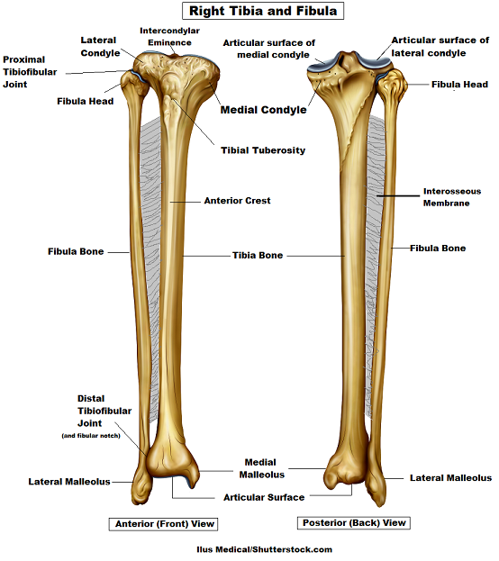
Leg Bones Diagram / How Equine Forelimb Anatomy Plays Out With Conformation And Soundness : The human leg consists of 8 bones, 4 per leg.. Anterior view with primary bones names. Quizzes on human skeletal system anatomy, bone anatomy, and bone markings. These can include any the following: Click now to learn more about the bones, muscles, and soft tissues tibia: Its lower end helps create the knee joint.
At the same time, the bones and joints of the leg and foot must be strong enough to support the body's weight while remaining flexible enough for movement and balance. Vector illustration with human skeleton scheme isolated on a white background. The largest and most medial leg bone, forming both the knee and ankle joints. Bones diagram human body 12 photos of the bones diagram human body all bones human body diagram, anatomy diagram human body. He'll boost his body knowledge as he matches up the names of the bones with their proper places on the leg diagram.
Tibia And Fibula Bone Anatomy from www.registerednursern.com He'll boost his body knowledge as he matches up the names of the bones with their proper places on the leg diagram. Quizzes on human skeletal system anatomy, bone anatomy, and bone markings. Click now to learn more about the bones, muscles, and soft tissues tibia: Learn how to draw the femur, patella, tibia, and fibula in this lesson! Diagram of blood and nerve supply to bone. This bright worksheet helps your child bring these technical terms down to size. The bones of the leg are the femur, tibia, fibula and patella. High resolution textures and displacement included.
The femur, or thighbone, is the longest and largest bone in the human body.
Each leg is made up of four bones. Bones of the leg and foot, lower leg bone anatomy, leg bones anatomy, leg muscles, leg bones diagram, leg bone structure, leg anatomy muscles, parts of the lower leg. Time to jump right into the biggest and strongest bones in the human body. Quizzes on human skeletal system anatomy, bone anatomy, and bone markings. These can include any the following: License image the bones of the leg are the femur, tibia, fibula and patella. Download the free graphic resources in the form of png, eps. Skeleton leg ankle joints and toe phalanges, cuboid, metatarsal, navicular and cuneiform bones, hand drawn dorsal view of foot. However, the definition in human anatomy refers only to the section of the lower limb extending from the knee to. Vector illustration with human skeleton scheme isolated on a white background. Human foot bones anatomy sketch of orthopedics medicine. This bright worksheet helps your child bring these technical terms down to size. Includes leg (femur, tibia, patella, and fibula) and foot (tarsals and digits) bones.
A leg bone is a bone found in the leg. Its lower end helps create the knee joint. Pngtree offers bone diagram png and vector images, as well as transparant background bone diagram clipart images and psd files. At the same time, the bones and joints of the leg and foot must be strong enough to support the body's weight while remaining flexible enough for movement and balance. Diagram of blood and nerve supply to bone.
Tibia And Fibula Bone Anatomy from www.registerednursern.com The foot bones shown in this diagram are the talus, navicular, cuneiform, cuboid. Blood vessels and nerves enter the bone. He leg's main function in the human is for locomotion and support of the rest of the body. Includes leg (femur, tibia, patella, and fibula) and foot (tarsals and digits) bones. High quality realistic skeleton legs. Your leg bones are the longest and strongest bones in your body. High resolution textures and displacement included. When you stand or walk, all the weight of your upper body rests on them.
At the same time, the bones and joints of the leg and foot must be strong enough to support the body's weight while remaining flexible enough for movement and balance.
Anterior view with primary bones names. The foot bones shown in this diagram are the talus, navicular, cuneiform, cuboid, metatarsals. At the microscopic level, this hard outer. These can include any the following: Your leg bones are the longest and strongest bones in your body. This bright worksheet helps your child bring these technical terms down to size. The bones involved in it, however, are only the femur and the tibia, although the smaller bone of the leg, the fibula, is carried along in the movements of flexion, extension, and slight rotation that this joint. At the same time, the bones and joints of the leg and foot must be strong enough to support the body's weight while remaining flexible enough for movement and balance. Use the leg bones diagrams to learn the names of the leg bones. Related posts of bones leg diagram picture. However, the definition in human anatomy refers only to the section of the lower limb extending from the knee to. Skeleton leg ankle joints and toe phalanges, cuboid, metatarsal, navicular and cuneiform bones, hand drawn dorsal view of foot. The musculoskeletal segment of the leg, including the foot bones (ankle, heel bone, toe bones), fibula and tibia, knee, femur and femoral neck, hip and sacrum as well as the third, fourth.
The bones involved in it, however, are only the femur and the tibia, although the smaller bone of the leg, the fibula, is carried along in the movements of flexion, extension, and slight rotation that this joint. At the same time, the bones and joints of the leg and foot must be strong enough to support the body's weight while remaining flexible enough for movement and balance. The foot bones shown in this diagram are the talus, navicular, cuneiform, cuboid, metatarsals. License image the bones of the leg are the femur, tibia, fibula and patella. The musculoskeletal segment of the leg, including the foot bones (ankle, heel bone, toe bones), fibula and tibia, knee, femur and femoral neck, hip and sacrum as well as the third, fourth.
Legs Bones Diagram Wiring Diagram Home Dress Improve Dress Improve Adcrent It from fpnotebook.com Bones diagram human body 12 photos of the bones diagram human body all bones human body diagram, anatomy diagram human body. Use the leg bones diagrams to learn the names of the leg bones. At the same time, the bones and joints of the leg and foot must be strong enough to support the body's weight while remaining flexible enough for movement and balance. Health diagram bone skeleton leg knee science anchor chart human human body. These simple labelled diagrams of the bones of the lower legs and feet and the bones of the arms and hands this diagram shows the skeletal structure of the leg (anterior view) and foot (dorsal view). The bones of the leg are the femur, tibia, fibula and patella. Bones of the leg and foot, lower leg bone anatomy, leg bones anatomy, leg muscles, leg bones diagram, leg bone structure, leg anatomy muscles, parts of the lower leg. Quizzes on human skeletal system anatomy, bone anatomy, and bone markings.
Your leg bones are the longest and strongest bones in your body.
Master leg and knee anatomy using our topic page. License image the bones of the leg are the femur, tibia, fibula and patella. Quizzes on human skeletal system anatomy, bone anatomy, and bone markings. When you stand or walk, all the weight of your upper body rests on them. Most bones (particularly the long bones of the arms and legs — which make up the appendicular skeleton) have a hard outer shell known as cortical bone. Blood vessels and nerves enter the bone. Visit kenhub for more skeletal system quizzes. These simple labelled diagrams of the bones of the lower legs and feet and the bones of the arms and hands this diagram shows the skeletal structure of the leg (anterior view) and foot (dorsal view). Each leg is made up of four bones. Bones diagram human body 12 photos of the bones diagram human body all bones human body diagram, anatomy diagram human body. Diagram of blood and nerve supply to bone. Learn how to draw the femur, patella, tibia, and fibula in this lesson! Human foot bones anatomy sketch of orthopedics medicine.

comment 0 komentar
more_vert